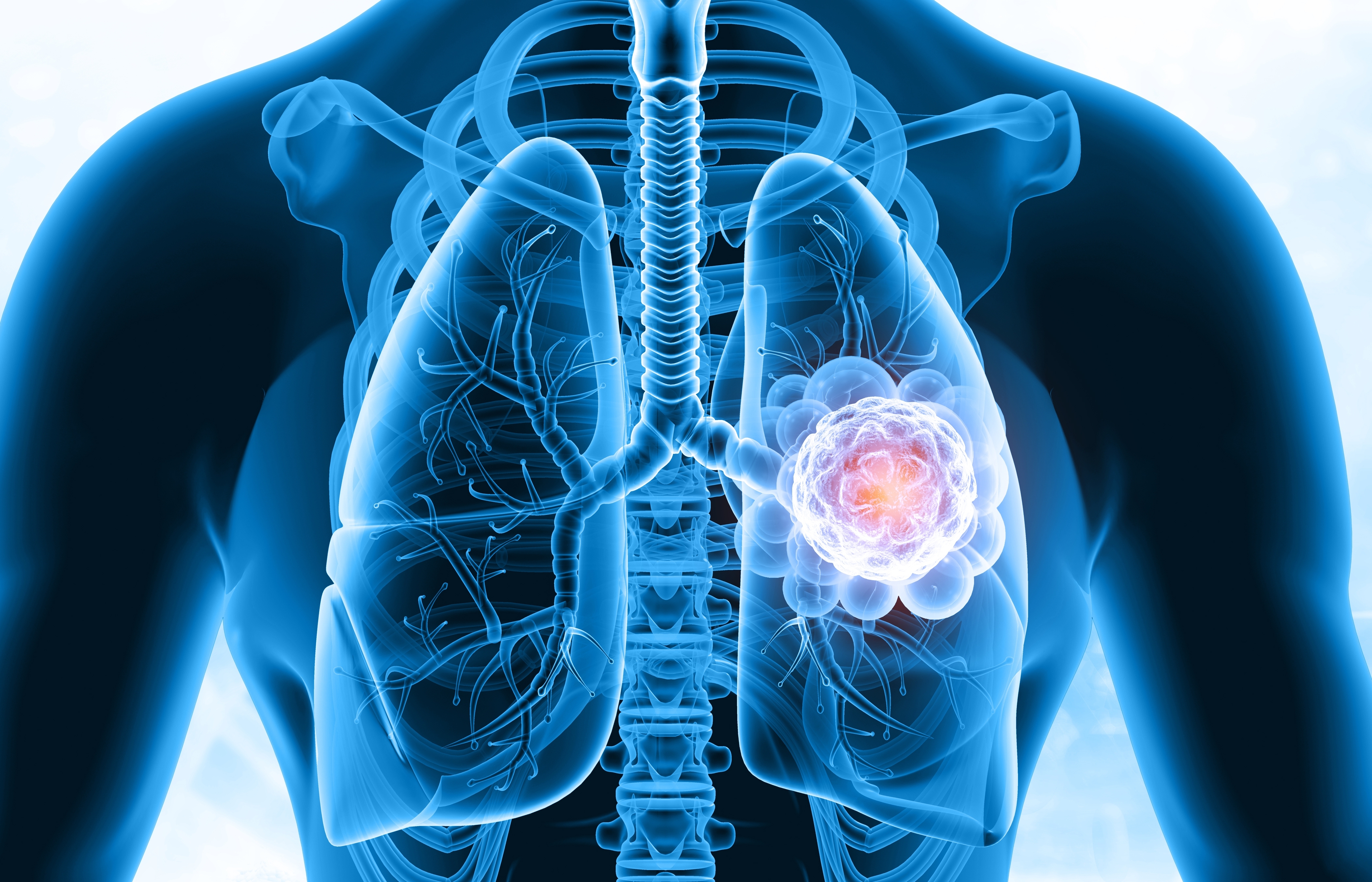
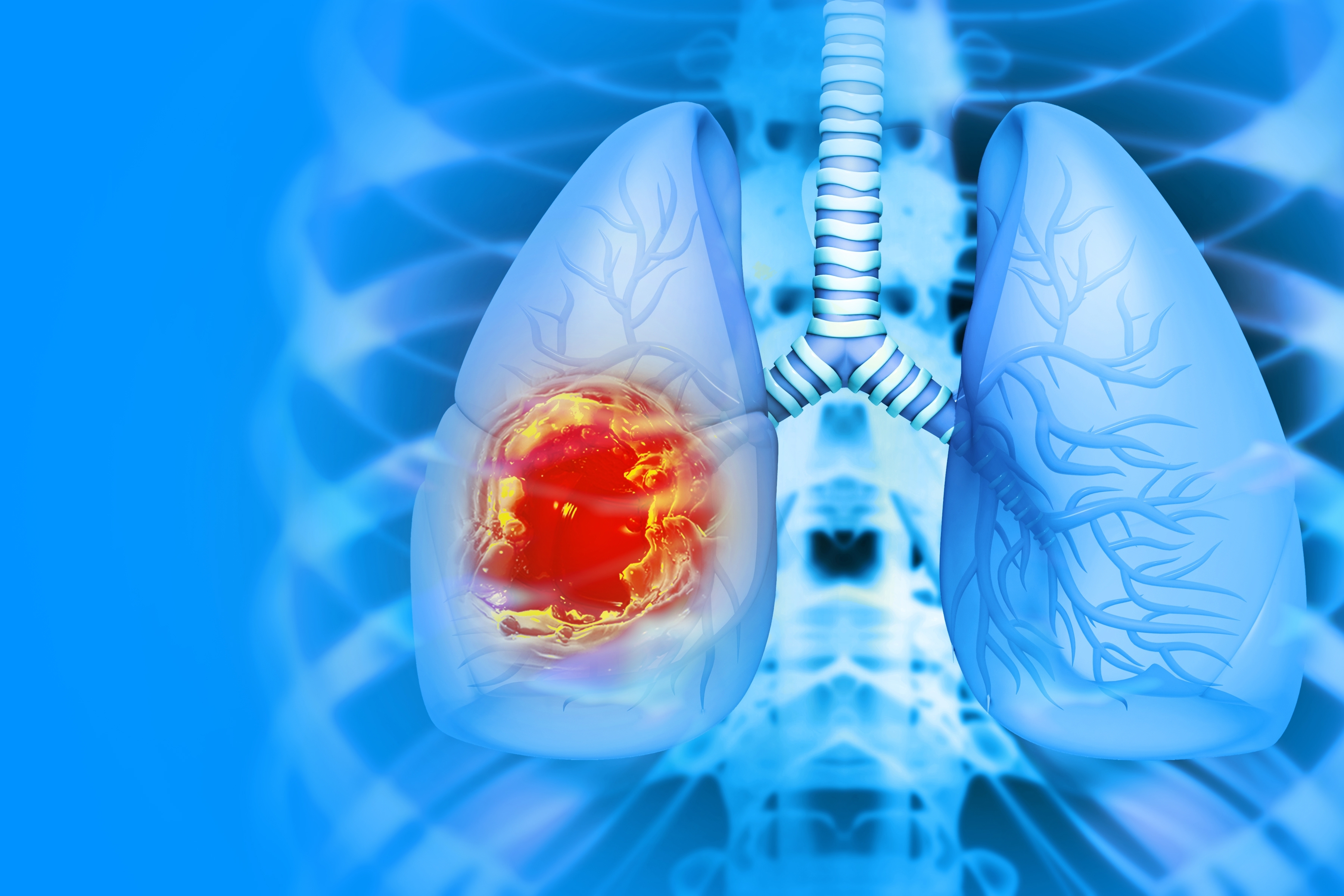
As a result of long and laborious Research and Development studies, Computer Aided Diagnosis System in Lung X-Ray Images have been developed for the first time in Turkey, which improves the quality of healthcare services by enabling the early detection of cancer.
Computer Aided Diagnosis System in Lung X-Ray Images
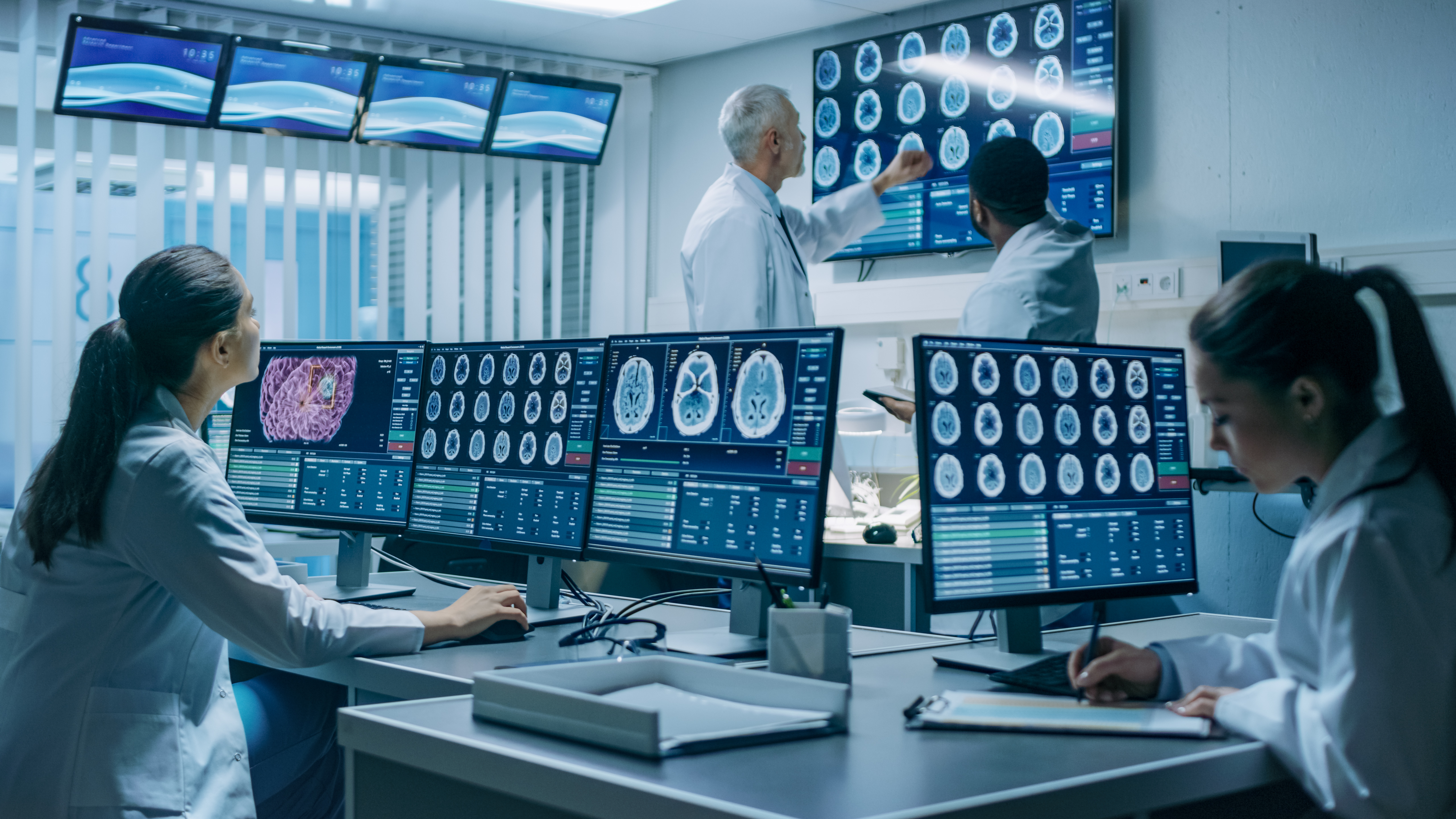
The software system determines the areas (target diagnosis or pathology) that are not obvious to radiologists in digital radiology images by using image processing and artificial intelligence methods. CAD is a secondaryand assistive diagnostic method while final decision belongs to the radiologist.
System Benefits
- To assist early diagnosis of lung cancer,
- To detect anomalies that may be missed by radiologists,
- To reduce misdiagnosis rate and increase accurate diagnosis rate,
- To help early detection of lung cancer from X-ray images,
- To speed up the diagnostic process using computer power,
- To reduce the workload of radiologists despite the increasing demand.
System Specifications
Anomaly Detection
- Detecting nodules with a radius size as small as 2.5 mm,
- Radiologist can mark detected nodules as correct/incorrect,
- Radiologists can mark additional detected nodules,
- All marked nodules can be displayed and recorded automatically in the generated reports.
Reporting and System that Learns
The system continuously learns as more patient x-ray images are reported.
Flexible Licensing and Different Work Models
- Flexible Licensing and Different Work Models based on end user needs.
PACS System Independency
- It can be integrated with any PACS system since it supports the International Standards.
Support for Education
- It can be used as a training tool in Universities, Hospitals etc.